
Microscopic and Microbial Insights into the Stony Coral Tissue Loss Disease (SCTLD) Outbreak Across Multiple Coral Species on Florida’s Coral Reef
Executive Summary
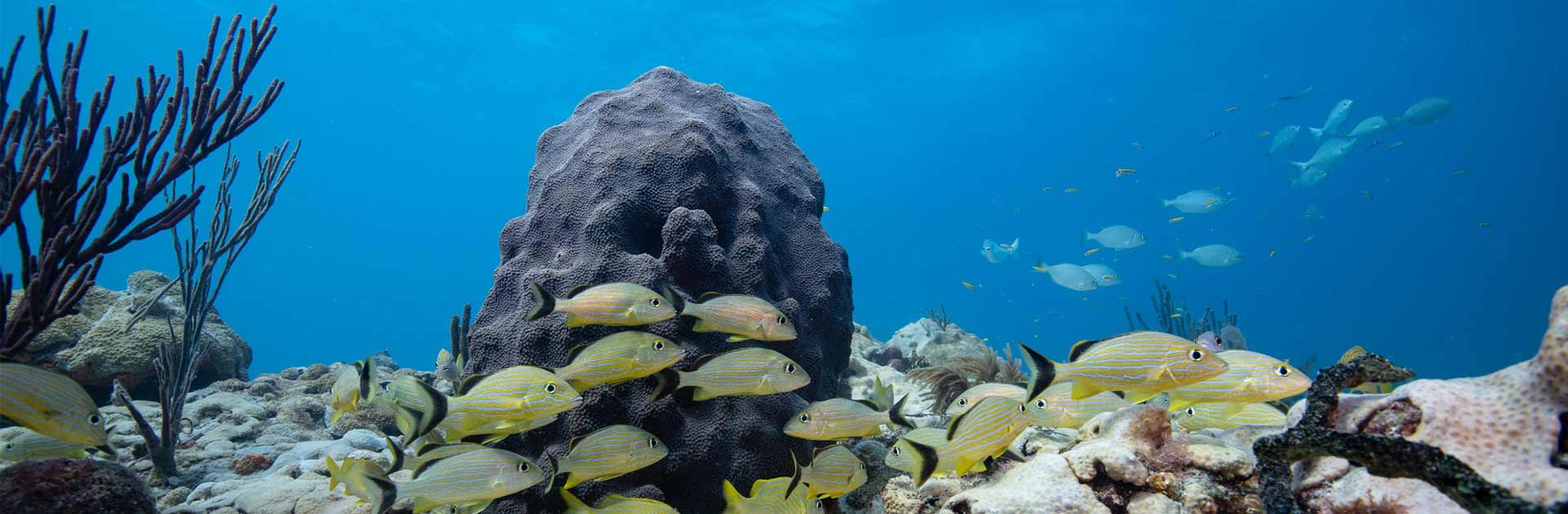
Three integrated research tasks were performed using molecular biology, histology, and immunohistochemistry to provide insights into a possible mechanism of bacterial infection that results in tissue loss, as well as potential pathogen(s) associated with stony coral tissue loss disease (SCTLD). For Task 1, molecular tools were used to characterize the endolithic communities of fungi, bacteria, and archaea from apparently healthy coral colonies, and affected and unaffected portions of diseased colonies sampled in 2016 and 2017. For Task 2 histological methods were used to characterize the endosymbiotic dinoflagellates in mucus and tissue from healthy, diseased, and unaffected portions of colonies and examine their role in the pathogenesis of SCTLD. For Task 3 immunohistochemistry was used to investigate the role of programmed cell death (PCD) vs. necrosis in SCTLD. Due to time constraints imposed by COVID-19- associated laboratory closures, further work is needed. Preliminary results from Task 1 revealed that the endolithic coral microbiome (bacteria and fungi) varied in community composition based primarily on host coral species and then disease state, especially regarding the fungal microbiome. Additional sample replicates and bioinformatic analyses will be performed to further investigate specific microbes driving these observed differences, as well as the functional potential of the coral microbiome during different disease states. Preliminary results from Task 2 showed that dinoflagellate abundance was reduced in the gastrodermis of most of the 2016 samples examined and remaining zooxanthellae were often degraded or lysing in the apparently healthy, diseased, and unaffected tissue samples from all four species of coral. Liquefactive necrosis was observed in all species, with few to no zooxanthellae in these lesions that were usually restricted to the gastrodermal tissue with the mesoglea and epidermis remaining intact. Gastrodermis adjacent to these lesions included palestinian algal cells with vacuolation, swelling, then cell wall disintegration and lysing (ghosting). Preliminary results from Task 3 revealed apoptosis was consistently observed in tissues of SCTLD-affected corals, indicating that this mode of cell death is involved in SCTLD pathology. However, apoptosis was also observed in some tissue without an apparent lesion, indicating that apoptosis may be an early indicator of disease before degradation can be detected morphologically. Additional samples and further analysis are needed to understand normal apoptosis involved in cell replacement and fully describe the interplay between the mechanism of cell death and other histological indicators of SCTLD. Continuation of this project will utilize the full sample set to better understand disease mechanisms and to identify potential pathogenic agents involved in the progression of SCTLD in reef-building corals.
Interested in subscribing to DEP newsletters or receiving DEP updates through email?
Sign UpThe Florida Department of Environmental Protection is the state’s lead agency for environmental management and stewardship – protecting our air, water and land. The vision of the Florida Department of Environmental Protection is to create strong community partnerships, safeguard Florida’s natural resources and enhance its ecosystems.
Learn MoreTallahassee, Florida 32399-3000 Public.Services@FloridaDEP.gov 850-245-2118
